Five Limits Your Brain Puts on Generosity
Research suggests that our brains may be wired for altruism, but there’s a catch—well, five of them, actually.
Humans can be remarkably generous.
Americans gave a record $390 billion to charitable organizations in 2016 through a combination of individual giving and philanthropy from estates, corporations, and foundations. And people give in myriad other ways as well, from everyday acts of kindness toward loved ones to volunteering to large acts of altruism, like donating a kidney to a stranger.
This isn’t surprising, given how wired we appear to be for giving.
But there are limits to our generosity—and many people want to be more generous than they actually are. We can all recall times when we refused to give to a person soliciting for a charity or failed to offer as much assistance to a friend or a stranger as we could have. If generosity feels rewarding to the giver as well as the recipient, what stops people from being generous to everyone all the time? Just as our brains have mechanisms in place that support generosity, studies in neuroscience have found ways that our brains rein in our generous tendencies.
Here are five that stand out.
1. Deliberation
We depend on our prefrontal cortex for many things—such as setting goals, creating plans, and making decisions—but work by UCLA researchers Leonardo Christov-Moore and Marco Iacoboni suggests that activity in parts of the prefrontal cortex can dampen our generous impulses in interesting ways.
In one study, the researchers used a technique called continuous theta burst stimulation (TBS) to disrupt the activity of one of two parts of the prefrontal cortex—the right dorsolateral prefrontal cortex (DLPFC) or the dorsomedial prefrontal cortex (DMPFC)—in two groups of participants. As a control, another group of participants received TBS in a brain region involved in perceiving movement.
While parts of their brains were still impaired by TBS, participants played something called a dictator game to test their generosity. In each round of this game, participants were given $10 and asked how much of that $10 they would choose to keep and how much they would give to a stranger identified by a headshot, name, and income level. Participants were told that, for a random selection of rounds, real money would be distributed as they selected, and they played the dictator game anonymously to ensure they weren’t trying to impress the experimenters with their generosity.
The result? Disrupting the activity of either the DLPFC or the DMPFC made people more generous (disrupting the control area had no effect).
The researchers write, “This suggests that our primary drive in non-strategic social transactions may in fact be to behave prosocially, perhaps due to reflexive forms of empathy that blur the boundaries between individuals.” In other words, we may default toward generosity unless a managerial part of the brain overrides that default and tells us to be stingy.
Intriguingly, the two non-control groups in the study became more generous in different ways. DLPFC disruption led people to give more money to high-income people compared to people who had a non-disrupted DLPFC. Disrupting the DMPFC, on the other hand, made participants more generous to the low-income strangers.
According to the researchers, these findings suggest that both the DLPFC and the DMPFC act to inhibit our inherent tendency to behave in ways that benefit others. Specifically, they suggest that activity in the DMPFC may act as a form of tonic control—a general stinginess signal—while the DLPFC responds more to context—perhaps persuading us to consider who could really use our generosity.
2. Lack of “neural empathy”
Another recent study by Christov-Moore and Iacoboni found evidence of another way our brains limit generosity: by inhibiting our “neural empathy.” Neural empathy is when we see another person in pain or expressing an emotion and parts of our brain process this experience as if we too were actually feeling the pain or emotion.
Using functional magnetic resonance imaging (fMRI), the researchers measured “self-other resonance,” a hallmark of neural empathy, in the brains of 20 participants while the participants watched three videos: one of a human hand alone, one of a hand being pierced with a hypodermic syringe, and one of a hand being touched by a Q-tip. While in the scanner, the participants also viewed or imitated photos of people making facial expressions. Outside the scanner, the participants played a dictator game to test their generosity.
Christov-Moore and Iacoboni found that participants who showed greater signs of neural empathy in the brain-imaging part of the study tended to be more generous while playing the dictator game.
For example, during the facial emotion mimicking task, the participants with more activity in their left amygdala—an area associated with neural resonance—and their left fusiform cortex—an area associated with empathy—gave more money to strangers with low incomes compared to people with lower activity in those areas.
Neural empathy is not the end of the story, however.
3. Prejudice
How our brains respond to another person’s emotions or pain can be influenced by a host of factors, including how well we know them, and whether or not they share our favorite soccer team, socioeconomic status, religion, and—perhaps most perniciously—race.
A number of studies have found that when a person observes another person in pain, there is more activity in the brain regions involved in perceiving this pain when both people share the same ethnicity or race.
One recent study suggests that this racial bias for neural empathy may show on our faces—literally. Shihui Han and colleagues at Peking University used electroencephalography (EEG) to record the brain activity of 24 Chinese college students while they viewed photos of Asian and European faces displaying neutral or pained expressions.
The participants showed significantly more activity in one type of brain wave, called N1, when participants viewed the pained expressions versus the neutral expressions, indicating that the pained pictures induced neural empathy. Notably, though, this effect was stronger when the participant and the person in the photo shared the same race.
Additionally, the increased neural empathy was largely blocked when a participant had a pen in their mouth, suggesting that facial mimicry plays an important role in processing other people’s emotions. However, the effect was not seen for the photos of white people. This suggests that the participants’ brains processed the facial expressions of people from their racial in-group differently from those of people outside their race.
A follow-up study by the same group looked at the relationship between racial prejudice and empathic neural responses. Specifically, this study tested whether there is a connection between individuation bias—the tendency to perceive members of one’s own race as individuals while generalizing people from other races—and the automatic responses the brain makes when seeing people in pain.
Han and colleagues were particularly interested in two EEG measurements: the so-called N170 signal, which responds to individual faces, and the P2 signal, which responds when people view other people in pain.
The researchers found that participants showed stronger N170 signals when viewing pictures of people who shared their race compared to those who did not. They also had a smaller P2 response when viewing photos of people outside their race, suggesting that the participants had a harder time perceiving people of other races as individuals and also showed less neural empathy for them. Additionally, the people who scored the highest on a racial prejudice test had the strongest neural markers for individuation bias and had the smallest P2 empathy responses to other-race photos.
The authors write, “It seems that prejudice prevents people from allocating cognitive resources to individuating members of racial out-groups in the first place, which makes racial out-groups even less identifiable for the purpose of empathizing.” This could have real consequences for everything from racially biased pain treatments to criminal sentencing.
But if prejudice can inhibit neural empathy, does that mean that neural empathy can be changed? Can we make our neural empathy less biased? The answer is yes, of course. While some of the factors that underlie our neural empathy for people of other races may be hard to change—having a particular oxytocin receptor gene variant, for example—some studies have shown that neural empathy is malleable and can be shaped by a number of external factors.
For example, a study by Han and colleagues at Peking University found that significant real-life experience with people of other races can reduce the racial bias seen in the empathic responses to another person in pain. In this study, Chinese adults who grew up in countries populated mostly by Caucasians showed the same neural empathy in response to videos of white and Chinese people in pain.
This, along with other studies, suggests that interacting with people who are different from us may change our brain’s automatic neural empathy—and our generosity.
4. No identifiable victim
Empathy depends on a feeling of person-to-person connection. Several studies have found that people are less generous toward multiple or anonymous victims—even the victims of large-scale disasters in dire need of help—than they are toward one specific, identifiable person. This is called the “identifiable victim effect.”
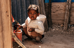
In one study, people were more likely to give money to another participant who had lost money in the experiment if that person was identified even by just a number rather than being completely unidentified. Another studyfound that people who saw a photo of a starving girl and read a description of her gave more money to an anti-hunger charity than did people who read statistics about starvation in Africa. And yet another study found that people were most likely to donate money for a sick child’s medical care when presented with the child’s name, age, and photo rather than just an age or an age and a name.
But why are we stingier with an anonymous potential aid recipient than we are with an identifiable person, even when we understand both may need our help?
A study by Alexander Genevsky and Brian Knutson and colleagues at Stanford and the University of Oregon explored this question. The researchers did this by giving undergraduate and graduate students $15 and then soliciting them for charitable donations while scanning their brain activity. Following their giving decision, the participants also reported how positive or negative they felt during the solicitation/donation scenario as well as their level of emotional arousal.
The researchers found that the students gave more money to orphans depicted by photographs than those shown as silhouettes. Interestingly, this study did not find that including a victim’s name increased donations or positive emotional arousal.
While a number of brain regions were more active when people were looking at a photograph than a silhouette, only activity in one brain region—the nucleus accumbens, a structure located toward the middle of the brain that is involved in motivation and reward—could account for the increased donations in the photograph scenario.
Besides providing a neuroanatomical basis for the identifiable victim effect, this study also provides insight into the possible role of emotional arousal in generosity. Crucially, the researchers found that seeing a photograph of an orphan led people to feel more positive emotional arousal than they felt while seeing a silhouette. This, in turn, led them to donate more. Negative arousal—as one may feel when experiencing guilt, for example—actually decreased giving.
Overall, this study suggests that information about a potential charity that increases positive emotional arousal—be it a photograph, a story, or some other information—may also increase generosity.
5. Adolescence
A new study from the Université Laval in Québec, Canada, suggests that teens may have less altruistic motivation to help others compared to adults, in part because their brains respond differently to people in need.
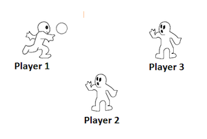
The researchers used fMRI to record the brain activity of twenty 12-17 year olds and twenty 22-30 year olds while they played a computer ball-tossing game called Cyberball, which simulates a social exclusion scenario.
Participants were led to believe that they would be playing Cyberball with other participants of the same age and were provided with pictures and names of these players. (In reality, the game was rigged by the experimenters). Players alternated between blocks of the game when they observed other players and blocks when they played themselves. Some of the observed rounds were manipulated so that one player was purposefully excluded and didn’t receive any of the throws. In the next round, study participants were given the opportunity to help the excluded player by including them in the game. This was how the researchers measured the players’ altruistic (or not so altruistic) tendencies.
Adolescents were much less generous compared with the adults. In particular, the mean number of throws to the excluded player was higher for the adult participants compared to the adolescents. Also, the adults gave a significantly higher proportion of their throws to the excluded players, at the expense of throws to the people who had done the excluding. The adolescents, however, did not show a significant difference in throws to the two groups.
This less helpful behavior in teens was underpinned by lower activity in several brain regions: the right temporoparietal junction, the fusiform face area, and the medial/dorsomedial prefrontal cortex. (Yes, activity in the prefrontal cortex was found to suppress generosity in a study mentioned above—our brains are complicated!).
Because the right temporoparietal junction and medial/dorsalmedial prefrontal cortex have been found to be active in experiments that ask participants to consider the mental states and perspectives of others, the researchers suggest that this lower activity level could be a possible cause of the less generous behavior in the teens. And, in fact, teens—on average—scored lower on a survey of perspective-taking in this study.
Importantly, older adolescents helped more compared to younger adolescents, suggesting that brain development may help explain the youngsters’ less generous behavior. And the researchers note that there could have been larger differences if they had compared the adolescents to older adults as there is evidence of some aspects of brain development continuing until age 30. So, if it seems as if your teen is not quite as helpful or generous as you were hoping, just take heart and wait a few years—this behavior may be the result of a still-developing brain.
Together, these studies show us various ways our brains limit generosity in different situations (and at different ages). Although we may think of generosity and altruism as virtues to aspire to, it makes a certain amount of sense that our brains have evolved to set limits. Without limits on generosity, we might deprive ourselves of the basic resources we need to function and thrive. We should all be glad for the limits our brains place on generosity, while simultaneously being aware of these limits so we can work to make sure we’re being our best, most generous selves.